Quick Answer: If you are preparing a neuroradiology talk — a brain tumor imaging lecture, an MR spectroscopy overview, a case conference for radiology residents — ChatSlide turns your topic, papers, and old decks into a complete slide deck in minutes. It is used by 260,000+ users across 750+ universities, searches PubMed and Google Scholar from inside the editor, builds full-length lecture decks with citations, generates charts from real data, writes per-slide speaker notes, and exports to editable PowerPoint on paid plans. Free to start, no card required.
The Neuroradiology Teaching Problem
Neuroradiology teaching is uniquely slide-hungry. A single brain tumor lecture has to move through sequence physics (why FLAIR, why DWI, why susceptibility), compartment localization, enhancement patterns, advanced techniques like perfusion and MR spectroscopy, and the molecular era of tumor classification — and every step wants an annotated image, a comparison table, or an algorithm figure. A neuroradiology fellow building a didactic, an attending preparing tumor board teaching, and a resident assembling their first case conference all hit the same wall: the interpretive expertise is the fast part, and the deck production is the slow one.
The result is familiar: talks assembled at midnight from screenshot fragments, citation lists that never quite match the slides, and a format that gets rebuilt from scratch for every rotation.
What Makes ChatSlide Powerful for Neuroradiology
PubMed and Google Scholar Inside the Editor
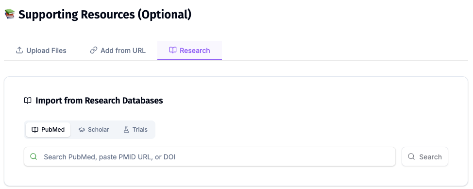
Neuroimaging evidence moves quickly — tumor classification updates, advanced-sequence validation studies, AI-in-radiology literature. ChatSlide's Research tab searches PubMed by keyword, PMID, or DOI and pulls key findings into your slides with citations, so the claim and its reference stay together. For techniques with pivotal trials still enrolling, the Clinical Trials search pulls design and endpoints by NCT number.
Case-Based Slide Generation
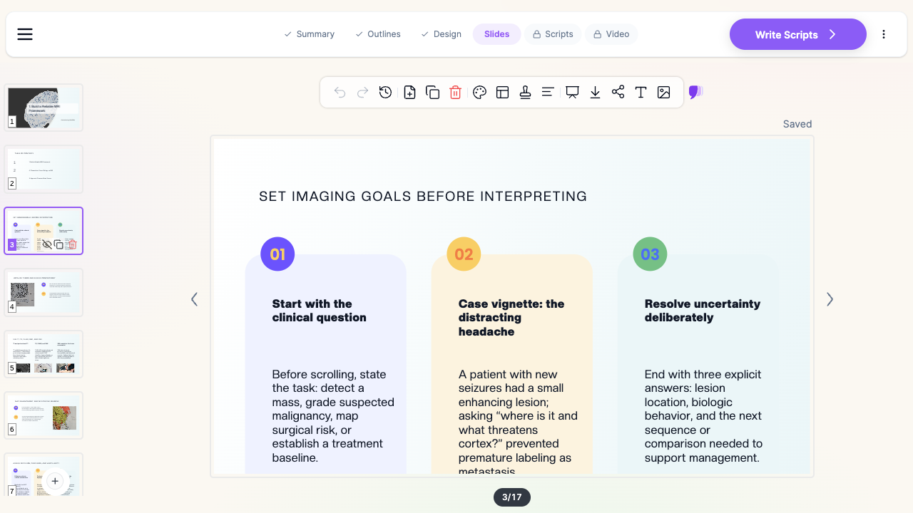
Describe a teaching case — "a patient with new seizures and a small enhancing cortical lesion" — and ChatSlide structures the deck the way neuroradiology teaching actually runs: clinical question, sequence selection, localization, signal characterization, differential, and the next study that resolves the uncertainty. The case thread becomes the spine of the talk instead of an afterthought.
Import Papers, Protocols, and Old Decks
Most neuroradiology talks start from something: a review article PDF, a departmental protocol document, last year's lecture. Upload PowerPoint or PDF files and ChatSlide extracts the content — including text locked inside figures, via OCR — and rebuilds it into a clean deck you update rather than recreate.
Your Images Stay Yours to Place
Generated imagery is a scaffold, not a substitute for cases. Drop your own de-identified DICOM exports, annotated key images, and spectroscopy curves into the generated layout — the deck structure, headings, and teaching text are already in place, so your time goes into the images that carry the teaching point.
Real Data Charts
Sequence-comparison tables, apparent diffusion coefficient ranges, metabolite ratios across lesion types — neuroimaging audiences expect data presented as data. Paste or upload your numbers and ChatSlide renders native editable charts instead of screenshots cropped from papers.
Speaker Notes You Can Teach From
Every slide gets AI-written speaker notes matched to its content. For an hour-long didactic, that is the difference between memorizing the deck and walking in with a script you have personalized with your own cases and pearls.
19 AI Editing Tools
Rewrite a physics slide for medical students, expand a bullet into a teaching paragraph, simplify fellow-level content for a mixed audience, translate for an international course — without breaking the deck's design.
How ChatSlide Builds Your Neuroradiology Deck
- Describe the talk or upload your sources. Type the topic and audience — "MRI of brain tumors and MR spectroscopy, for radiology residents" — or upload papers, protocols, and an old deck. ChatSlide extracts and organizes the content.
- Review the outline. The AI proposes a section-by-section structure — reorder, merge, or add sections to match your learning objectives.
- Generate slides with images and charts. ChatSlide writes the slides, adds supporting imagery, renders your data as charts, and attaches citations from the Research tab. Swap in your own annotated cases where they carry the teaching point.
- Polish and export. Edit anything inline, generate speaker notes, then export to PowerPoint or PDF — or record the deck as a narrated video.
Use Cases for Neuroradiologists and Trainees
The brain tumor imaging lecture. The core subspecialty didactic: compartment localization, enhancement patterns, diffusion and perfusion integration, and how imaging fits alongside histology and molecular classification. Generated as a structured lecture, then anchored with your own annotated cases.
The MR spectroscopy overview. A technique talk that has to teach metabolite peaks, acquisition choices, and — most importantly — where spectroscopy genuinely changes management versus where it adds noise. ChatSlide builds the didactic scaffold; your spectra make it real.
The resident case conference. A resident presenting unknowns needs case timeline slides, a differential that narrows visibly, and a reveal structure. Describe the case flow and the deck comes back organized for stepwise teaching.
Tumor board teaching. The five-minute imaging summary for a multidisciplinary audience: what the imaging shows, what it favors, and what it cannot answer — simplified from the same material with the rewrite tools.
The journal club or technique update. New sequence validation, AI triage tools, guideline updates — pull the papers through the Research tab and present the evidence with citations on the slides where the claims appear.
Teaching files for the rotation. Convert a folder of teaching cases into a consistent deck format once, then regenerate and extend it each rotation instead of rebuilding.
Neuroradiology AI Tools Compared (2026)
| Capability | ChatSlide | Gamma | Canva | Beautiful.ai |
|---|---|---|---|---|
PubMed / Scholar / NCT search with citations | ✅ Built-in | ❌ | ❌ | ❌ |
Full-length lecture decks | ✅ | ✅ (paid) | ✅ | ✅ |
PDF / PPT import with OCR | ✅ | Partial | Partial | Partial |
Editable data charts | ✅ | ✅ | ✅ | ✅ |
AI per-slide speaker notes | ✅ | ✅ | ✅ | ✅ |
AI-narrated video export | ✅ | ❌ | Partial | ❌ |
Editable PowerPoint export | ✅ (paid plans) | ✅ | ✅ | ✅ |
Free tier | ✅ | ✅ | ✅ | Trial only |
General-purpose deck tools produce attractive slides, but none connect to the research databases a subspecialty talk depends on — so the slowest part of the job, evidence and citations, stays manual.
Time Comparison: Manual vs. AI-Assisted
Typical estimates for a one-hour neuroradiology didactic — your numbers will vary with topic familiarity and how many of your own cases you place:
| Task | Manual | With ChatSlide |
|---|---|---|
Literature search and citation formatting | 3–5 hours | 30–45 min (Research tab, inline citations) |
Outline and case structure | 1–2 hours | Minutes (generated, then edited) |
Building 25–40 slides with layout and imagery | 5–8 hours | 15–30 min generation + 1–2 hours placing cases |
Charts and comparison tables | 1–2 hours | Minutes per chart |
Speaker notes | 2–3 hours | Generated, then personalized |
Total | 12–20 hours | 2–4 hours |
What a Strong Neuroradiology Talk Includes
The domain content still has to be right — AI accelerates the slides, not the interpretive teaching. A talk peers take seriously typically covers:
- The clinical question first. State what the study was asked to answer before showing a single image — detection, characterization, surgical planning, or treatment response — and return to it at the end.
- Sequence logic, not sequence lists. Teach why each sequence is in the protocol and what it uniquely contributes, rather than reciting the acquisition list.
- Localization as a discipline. Compartment and relationship to eloquent structures decided explicitly, with the findings that argue each way shown side by side.
- Advanced techniques with their limits stated. Diffusion, perfusion, and spectroscopy presented with the overlap and pitfalls acknowledged — including where they disagree and how to resolve it.
- The molecular context. Imaging conclusions framed alongside integrated histologic and molecular classification, with the discordant-case caveat made explicit.
- A case thread. Radiologists learn pattern plus process through patients; carry one or two de-identified cases through the deck.
If you are preparing a general radiology talk — department conferences, teaching files across body systems, QA presentations — see the radiology & medical imaging guide instead; this page is about the neuroimaging subspecialty. For clinical neurology talks built around management rather than imaging, see the neurology presentation guide.
Direct Research Database Access
ChatSlide's Research tab connects to the databases physicians use daily:
- PubMed: Search by keyword, PMID, or DOI. Find the landmark trials, recent publications, and clinical guidelines relevant to your case. The AI reads abstracts and incorporates key findings into your slides with citations.
- Google Scholar: When your topic spans disciplines — say, the intersection of genetics and oncology — Scholar captures the broader academic literature that PubMed alone might miss.
- Clinical Trials (NCT): Presenting on a treatment where pivotal trials are ongoing? Search by NCT number or condition to pull trial design, endpoints, and status into your slides.
Best Practices for Neuroradiology Slide Decks
Do:
- Anchor every technique or accuracy claim to a citation on the slide where it appears
- Annotate your images — an arrow and a one-line label beat a paragraph beside an unmarked image
- Show sequences side by side when the teaching point is their disagreement
- Carry a de-identified case through the talk as the teaching spine
- Write speaker notes as spoken language, not slide text repeated
Don't:
- Paste figure screenshots from papers when an editable chart is clearer
- Exceed roughly one slide per minute of talk time
- Present advanced-technique data without stating the pitfalls and overlap
- Put real patient data on slides — de-identify everything, including burned-in annotations on image exports, always
Note on patient data: ChatSlide's standard plans are not a HIPAA-covered service — keep PHI out of slide content and uploads. For hospital systems, group practices, and clinics that need a Business Associate Agreement, our Enterprise plan offers HIPAA-compliant deployment options — contact us to discuss BAA terms, SSO, and on-prem / private-cloud hosting.
ChatSlide for Radiology Departments and Fellowship Programs
Radiology departments, neuroradiology fellowship programs, and hospital education committees can standardize the pipeline: shared brand templates so every presenter's deck matches, team billing, SSO, and collaborative editing across sites. For organizations with compliance requirements, Enterprise deployment adds the BAA and private hosting options above — contact us to set up a team workspace.
Frequently Asked Questions
Can ChatSlide build a full neuroradiology didactic? Yes. Specify the length and structure in your prompt (sections, slides per section) and the generator builds the full deck; you then edit rather than author. Review the outline carefully before generating slides.
Can it include my own MRI images? Yes — and it should. Generated imagery scaffolds the deck, but the teaching cases are yours: upload de-identified image exports and place them on the generated slides. The import tools also pull content out of existing case-based decks, with OCR for text inside figures.
Does it cite real papers? The Research tab pulls real, verifiable entries from PubMed, Google Scholar, and ClinicalTrials.gov, and citations added from it link to the actual source. As with any tool — and any human-built deck — verify that each citation supports the specific claim before you teach it.
How is this different from the general radiology guide? The radiology & medical imaging guide covers department-level radiology talks across body systems. This page covers the neuroimaging subspecialty: brain tumor MRI, advanced sequences, spectroscopy, and the didactic, case-conference, and tumor-board formats that go with them.
Can it teach MR spectroscopy content correctly? It builds the structure and the didactic text; the spectra and metabolite interpretations should come from your own cases and the cited literature. That division — AI does the scaffold, you supply the subspecialty judgment — is the honest workflow, and the fastest one.
Can I turn a lecture into a video for asynchronous teaching? Yes. ChatSlide can narrate the deck with an AI voice or avatar presenter and export a video — common for fellowship curricula and resident teaching libraries.
Is my content used to train AI models? Your uploads and generated decks stay in your workspace. Organizations that need contractual guarantees on data handling — including training exclusions — get them in writing with the Enterprise plan: contact us.
How much does it cost? You can build your first decks free, with no card. Paid plans unlock PowerPoint export and lift limits on deck length, uploads, and video; Enterprise adds team and compliance features.
Get Started
Your next neuroradiology talk does not need to cost you a weekend. Make your neuroradiology presentation with ChatSlide — describe the case, pull the evidence from PubMed, place your images, and walk in with a deck, notes, and citations that look like they took a month.