Quick Answer: A teaching-quality sonography deck usually needs (1) an ultrasound physics primer — frequency vs penetration, axial/lateral resolution, harmonics, artifacts (shadowing, enhancement, reverberation), (2) knobology — gain, TGC, depth, focal zones, frequency, the freeze/measure workflow, (3) scanning protocol — patient prep, transducer selection, standardized views and required images per ARDMS/AIUM, (4) normal anatomy with sonographic correlation — organ-by-organ landmarks and measurements, (5) pathology recognition — the classic appearances (cholelithiasis with shadowing, hydronephrosis, appendicitis, free fluid), (6) Doppler — color, power, spectral, aliasing and the Nyquist limit, (7) artifact and pitfall slides so trainees don't over-call. Target 25–35 slides for a 60-minute class, 12–18 for an SPI board-review session. ChatSlide accepts AIUM practice parameters and protocol PDFs and produces the structured lecture format sonography programs expect.
The Sonography Lecture Tax
Diagnostic medical sonography is one of the most image-dependent things you can possibly teach, and that makes the slide deck punishing to build. A single class on the right upper quadrant exam pulls from an AIUM practice parameter, a protocol manual, a stack of teaching images, the SPI physics curriculum, and a decade of scans you've personally saved. Sonography students preparing for the ARDMS SPI (Sonography Principles & Instrumentation) exam, registered sonographers (RDMS/RVT/RDCS) chasing CME, and program faculty assembling a semester of didactics all hit the same wall: most of the content is settled, well-codified material — the resolution trade-offs, the standard views, the measurement criteria — but assembling the deck still eats the whole weekend.
Build it the old way and you spend Saturday hunting for a clean transducer diagram, redrawing the relationship between frequency and penetration, and copying organ measurement cutoffs out of a protocol manual. By Sunday afternoon the class is half-built and you're cropping images in Preview because PowerPoint mangled the aspect ratio on your liver montage.
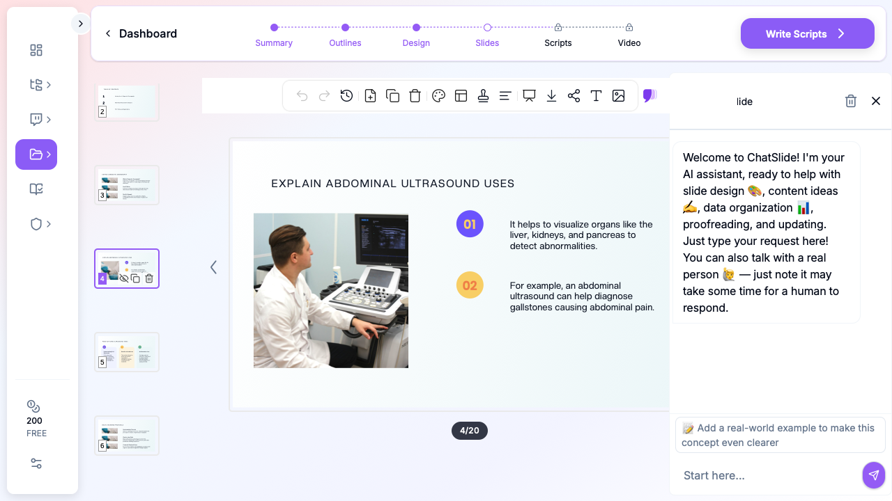
ChatSlide collapses the assembly step. Drop a topic — "Right upper quadrant abdominal ultrasound protocol for second-semester sonography students" or "Ultrasound physics and instrumentation for the ARDMS SPI exam" — and you get a structured lecture deck with a clear teaching arc, anatomy and equipment visuals, side-by-side comparison tables, and speaker-ready talking points. You stay in charge of the clinical judgement and the image curation; the tool stops eating your weekend.
What a Strong Sonography Lecture Actually Needs
Sonography audiences are unusually demanding because the same room often holds a first-semester student who has never held a transducer and a 20-year RDMS picking up a CME credit. A teaching-quality sonography deck has to hit a specific set of bars:
A physics primer that respects the SPI. The frequency-vs-penetration trade-off, axial vs lateral resolution, the focal zone, harmonic imaging, and the major artifacts (acoustic shadowing, posterior enhancement, reverberation, mirror image, edge artifact) are the spine of the SPI exam. Show the trade-off once, cleanly, on a single plate — four bullets re-reading a textbook lose the room immediately.
Knobology framed around problem-solving. Students don't struggle with the definition of time-gain compensation — they struggle with knowing which control fixes a too-dark far field. Open with a poor image and the two or three adjustments (gain, TGC sliders, depth, focal zone) that fix it. That single slide does more teaching than an entire physics chapter.
Scanning protocols that match the practice parameters. AIUM and the ARDMS body-of-knowledge define the required images for each exam — the RUQ study, the complete abdominal survey, the renal exam, the first- and second-trimester OB exams, the venous duplex. Your protocol slides need to match those required-image lists exactly, because that is what students are tested on and what labs audit against.
Normal anatomy with true sonographic correlation. A line drawing of the biliary tree is necessary but not sufficient — students need the drawing next to the actual sonographic appearance, with the common bile duct measured and the portal triad labeled. Echogenicity language ("hyperechoic," "anechoic," "complex") only sticks when it's anchored to a real image.
Pathology by its classic appearance. Cholelithiasis with clean posterior shadowing, hydronephrosis grading, the non-compressible blind-ending tube of appendicitis, free fluid in Morrison's pouch, the thickened gallbladder wall with pericholecystic fluid. Trainees memorize patterns; your slides should present each entity as a recognizable pattern, not a paragraph.
Doppler that connects to flow. Color vs power vs spectral Doppler, the angle-correction rule, and aliasing/Nyquist belong in any vascular or OB lecture. The moment a slide says "aliasing occurs when…" without a side-by-side tracing, the room tunes out.
Artifacts taught as friend and foe. Some artifacts are diagnostic (shadowing behind a stone, twinkle artifact on a calculus, comet-tail in adenomyomatosis); some cause errors (mirror image above the diaphragm, side-lobe pseudo-debris). A dedicated artifact slide prevents the most common student over-calls.
Real cases with annotated images. Three or four cases woven through the talk — normal, then a clear abnormal, then an ambiguous one — wake the room up far better than another physics slide.
Step-by-Step: Building Your Sonography Lecture with ChatSlide
1. Pick the right scenario
In ChatSlide, choose Education > Lecture for program didactics, SPI prep classes, or CME workshops. This biases the outline generator toward pedagogical structure — primer, principles, protocol, pathology, cases, summary — rather than the conference-keynote shape you'd want for an AIUM scientific session.
For a focused conference talk (say, "Contrast-enhanced ultrasound of focal liver lesions"), switch to Conference > Keynote. For a journal-club review of an ultrasound-endpoint trial, Education > Journal Club generates the critical-appraisal structure.
2. Write the topic the way you'd describe it to a colleague
Generic strings produce generic decks. Compare:
- Bad: "Ultrasound lecture"
- Better: "Abdominal ultrasound protocol for sonography students"
- Best: "Right upper quadrant and complete abdominal ultrasound protocol — transducer selection, required AIUM images, normal measurements, and biliary/renal pathology, for second-semester DMS students"
The third version gives the AI enough to build a focused 28-slide deck with the protocol alignment your students actually need.
3. Define the audience precisely
"Sonography students" and "experienced sonographers" produce different decks. Be explicit:
- "First-semester DMS students with no prior scanning experience"
- "Students preparing for the ARDMS SPI (Sonography Principles & Instrumentation) exam"
- "Registered sonographers (RDMS/RVT) earning CME on point-of-care abdominal ultrasound"
- "Emergency medicine residents learning the FAST and eFAST protocols"
The audience description changes the assumed baseline. A first-semester deck spends three minutes on transducer handling; a CME deck spends 30 seconds on it and moves to nuance.
4. Generate the outline first
Outline generation is fast and cheap. Read the section structure before generating slides. Common edits at this stage:
- Combine redundant primer sections (you don't need three slides on the piezoelectric effect)
- Add a "required images checklist" section if it didn't surface — students photograph these
- Reorder so the artifact slide comes after normal anatomy, not before
- For an SPI-prep deck, add a "high-yield physics pearls" closing section
5. Generate slides with images
This is where most generic AI presentation tools fall down. They produce text slides with stock-photo doctors holding clipboards. ChatSlide's image step pulls in transducer diagrams, anatomic illustrations, ultrasound-style cross-sections, and equipment visuals — the kind of imagery a sonography audience expects.
For modality-specific scans (sagittal RUQ, longitudinal kidney, the four FAST views, OB biometry), you'll want to swap in your own de-identified teaching clips. The deck structure stays intact — drag your captured images into the existing image slots. Patient identifiers and the burned-in banner should already be removed before you import; ChatSlide does not see your raw DICOMs or the machine archive.
6. Refine for the room
A 50-minute SPI review for fifteen students reads differently than a 90-minute hands-on CME workshop for forty working sonographers. Tighten the deck for the room:
- DMS program didactic: 25–35 slides, image-heavy, required-images checklist near the end
- ARDMS SPI board prep: 15–20 high-density physics slides, exam-style questions every 10 minutes
- Hands-on CME workshop: 20–25 slides, scanning-station references, less text per slide
- POCUS for EM/ICU residents: 12–15 slides, the FAST/eFAST views, IVC, lung B-lines, focused cardiac
- OB sonography module: 25–30 slides, first/second/third-trimester protocols and biometry
7. Export to PPTX or present from the browser
Exported PPTX preserves layout, bullets, and image positions — useful when the AV team in your lecture hall requires a local file. The browser preview also works well for small lab groups where you want to skip the export step and edit live.
Specific Use Cases This Replaces
Ultrasound physics modules. Frequency vs penetration, resolution, focal zones, harmonics, the artifact family — generate the SPI primer once, reuse for every incoming cohort.
Doppler comparison plates. Color vs power vs spectral, angle correction, aliasing and the Nyquist limit — the AI builds the side-by-side comparison; you verify the example tracings.
Protocol reference decks. RUQ, complete abdomen, renal, OB first/second/third trimester, venous duplex, thyroid, scrotal — generate one structured protocol deck per exam, link them as a series.
Pathology recognition teaching. Cholelithiasis, hydronephrosis, appendicitis, AAA, DVT, ectopic pregnancy — start a deck from the classic appearance and let the outline build the supporting differentials.
POCUS / FAST training. A focused deck for non-radiology trainees — the four FAST views, eFAST lung windows, IVC assessment — narrower, less physics, more "what does this view tell you in 10 seconds."
ARDMS / SPI board-review series. Build a multi-session series by listing all SPI topics, running each through ChatSlide, and linking the decks. Students get a consistent visual language across the whole review.
Direct Research Database Access
ChatSlide's Research tab connects to the databases physicians use daily:
- PubMed: Search by keyword, PMID, or DOI. Find the landmark trials, recent publications, and clinical guidelines relevant to your case. The AI reads abstracts and incorporates key findings into your slides with citations.
- Google Scholar: When your topic spans disciplines — say, the intersection of genetics and oncology — Scholar captures the broader academic literature that PubMed alone might miss.
- Clinical Trials (NCT): Presenting on a treatment where pivotal trials are ongoing? Search by NCT number or condition to pull trial design, endpoints, and status into your slides.
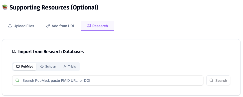
For sonography specifically, the Research tab is most useful for pulling in the AIUM practice parameters and consensus statements, the SRU (Society of Radiologists in Ultrasound) consensus criteria (for example, thyroid nodule and ovarian lesion management), and the diagnostic-accuracy studies that underpin POCUS protocols like the FAST exam and lung ultrasound.
Tips Specific to Sonography Lectures
Lead with knobology, not the piezoelectric effect. Students never struggle with the physics definition — they struggle with which control fixes the image. Open with a poor-quality scan and the adjustments that fix it.
Show the artifact, don't describe it. A shadowing stone, a posterior-enhancement cyst, a mirror-image diaphragm — two real images side-by-side teach more than four bullets explaining the acoustics.
One required-images checklist per protocol. Students will literally photograph the checklist slide. Make it match the ARDMS/AIUM required-image list exactly.
Anchor every echogenicity term to an image. "Hyperechoic," "anechoic," "complex," "isoechoic" only stick when each is shown on a real scan, not defined in prose.
Be explicit about technique limitations. Bowel gas obscuring the pancreas, body habitus and penetration, operator dependence — slides that name the limitations land harder than slides that pretend ultrasound always works.
End with high-yield SPI pearls. Students photograph the pearls slide. Make it clean, physics-aligned, and worth photographing.
Skip stock photos of doctors with clipboards. A deck full of staged photos signals you didn't put time into the talk. Transducer diagrams, real (de-identified) sonographic clips, and protocol schematics only.
Why Generic AI Presentation Tools Underperform Here
Most AI deck generators were built for sales pitches and marketing decks. They optimize for visual flair and ignore the structural conventions of clinical didactics. For a sonography audience, that produces:
- Physics slides with cartoon waves and no actual transducer cross-section
- Doppler slides that confuse color and spectral because the source didn't explain the difference
- Protocol slides that miss the required AIUM images
- Stock-photo "doctor reviewing chart" slides that have no place in an ultrasound class
ChatSlide's clinical scenarios — Education, Conference, Research, Journal Club — bias the output toward the conventions a sonography audience expects. You're still the clinical author; the tool just stops fighting your format.
Get Started
Building your next diagnostic medical sonography lecture on ChatSlide takes about as long as completing two studies. Drop a topic, refine the outline, generate the slides, swap in your own de-identified clips, and you have a class-ready deck before the next lab session.
Try it free at chatslide.ai — most sonography educators and POCUS instructors go from blank screen to first draft in under fifteen minutes.
Related guides:
- Echocardiography Presentation AI for Board Review
- Medical Imaging and Radiology Presentation AI Guide
- AI Medical Presentation Maker for Healthcare Professionals
- Medical Board Review Presentation AI Guide
- Patient Education Presentation AI Guide
A note on patient data and HIPAA. ChatSlide's standard plans are not a HIPAA-covered service — keep PHI out of slide content, prompts, and uploads. De-identify your ultrasound clips and case data — and strip the burned-in patient banner — before you import. For hospital systems, academic medical centers, and ultrasound/imaging departments that need a Business Associate Agreement, our Enterprise plan offers HIPAA-compliant deployment options — contact us to discuss BAA terms, SSO, and private-cloud or on-prem hosting.